How science turned back the clock of human development to create a miracle blood stem cell

Many thought it could never be done. But three Murdoch Children’s Research Institute scientists recreated embryonic development to crack the blood stem cell code.
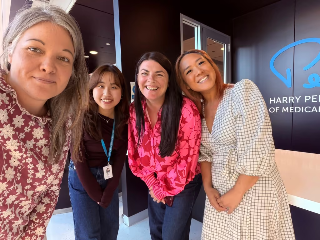
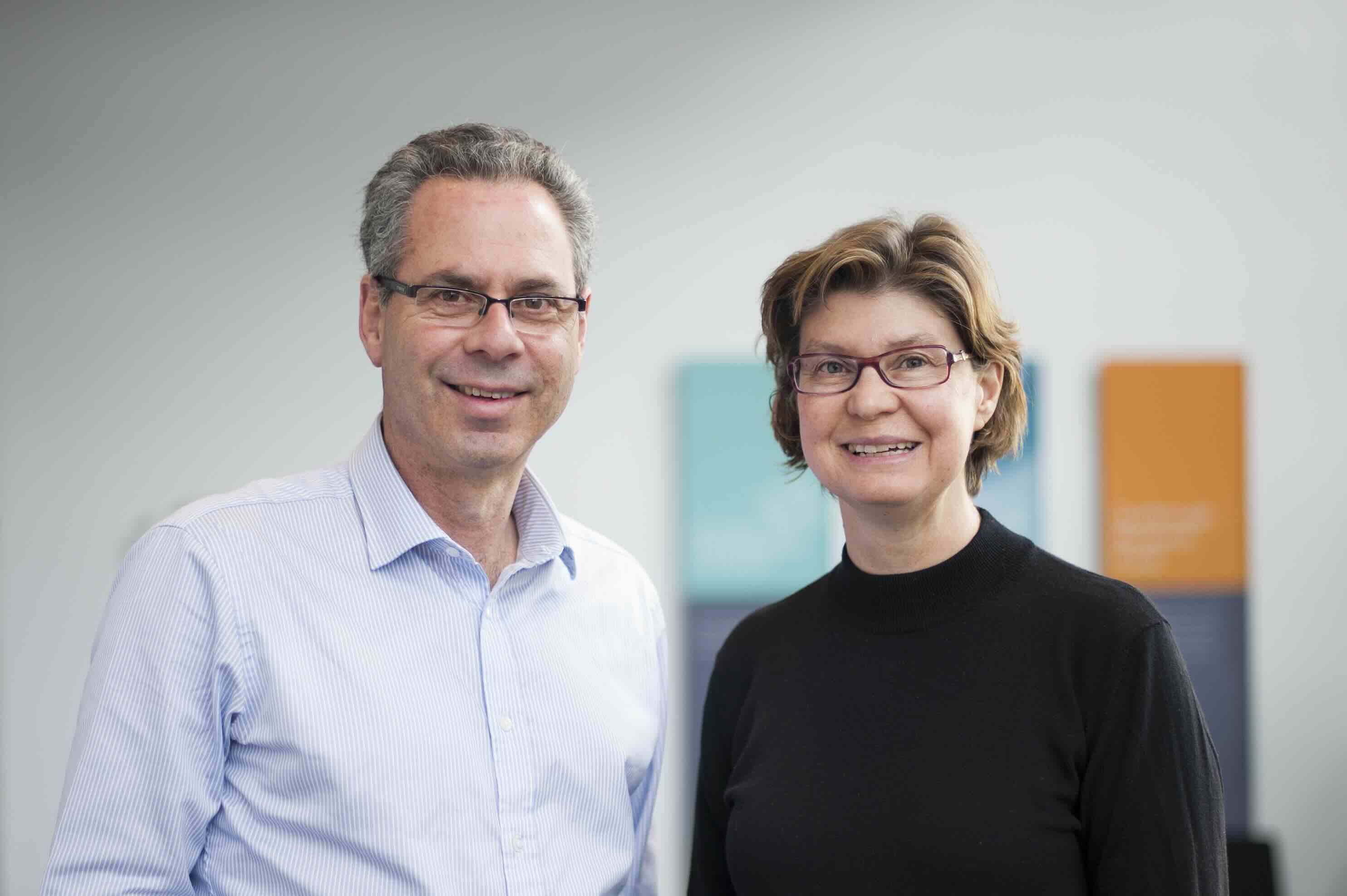
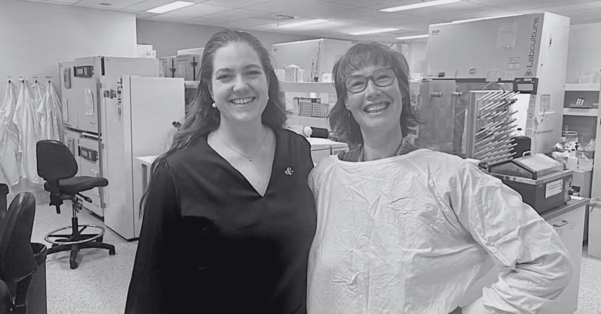
HM1 Chief Operating Officer Maggie O’Neill with A/Prof Elizabeth Ng at the Murdoch Children's Research Institute in Melbourne (March 2026).

Natasha Robinson

How science turned back the clock of human development to create a miracle blood stem cell

May 4, 2026
Many thought it could never be done. But three Murdoch Children’s Research Institute scientists recreated embryonic development to crack the blood stem cell code.
Read Transcript

Scientist Elizabeth Ng is clutching a small flask with a clear mixture inside it that she’s just retrieved from inside a bioreactor.

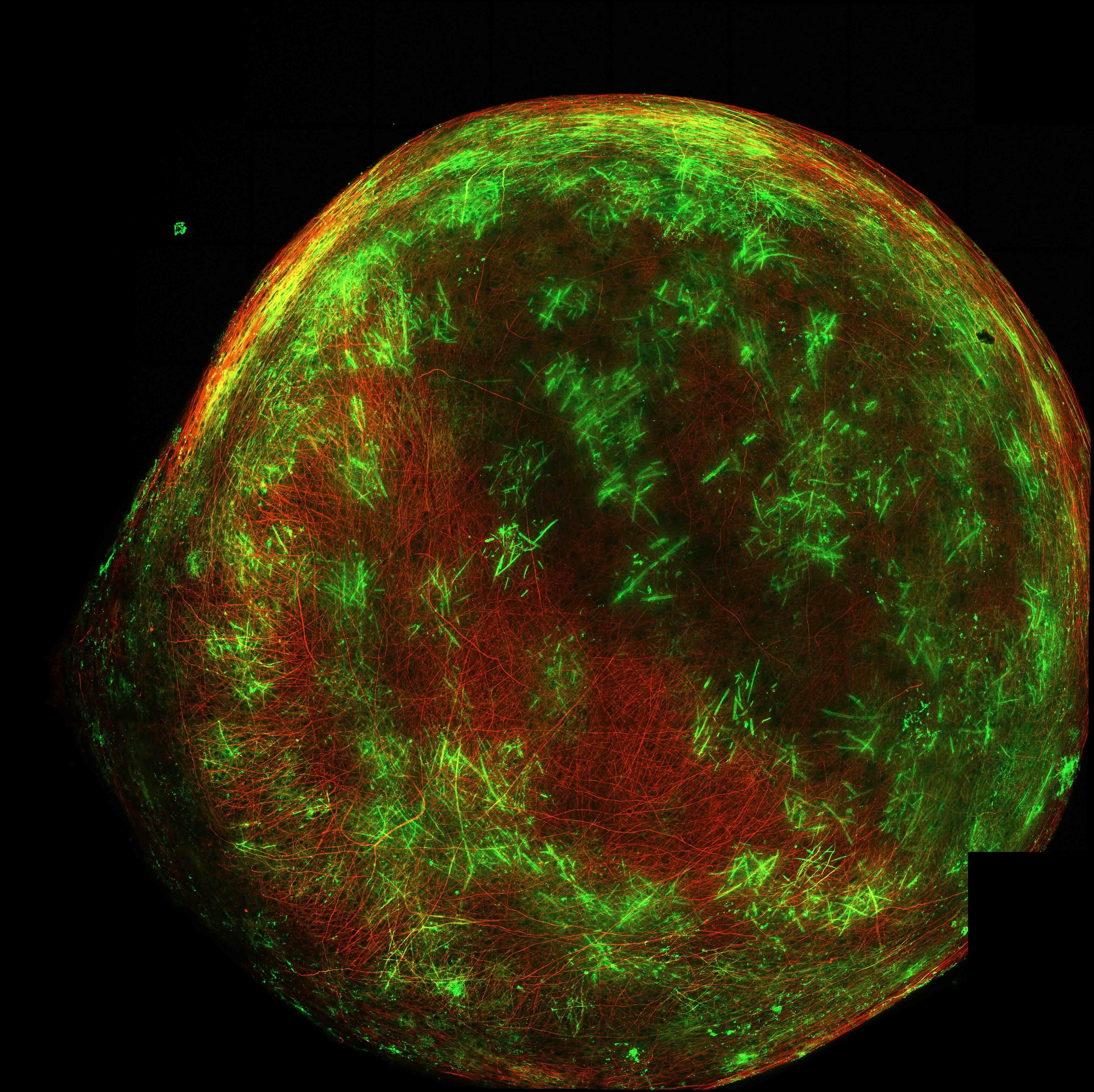
The colonies of cells clearly visible inside the flask are the genesis of life: pluripotent stem cells. They’re capable of forming every tissue in the body. Inside these flasks, the induced pluripotent stem cells are forming tiny clusters that replicate the human embryo in its earliest iteration.

“These pluripotent stem cells are immortal,” Associate Professor Ng says from inside the Blood Development Laboratory at the Murdoch Children’s Research Institute in Melbourne, where she is a group leader.

“We can use them to understand how to make human tissues. To do that, we have to recreate every step in early embryonic development. We have to deconstruct that process, and then mimic the whole process from scratch in the laboratory.”

This is the incredible story of how three Australian scientists, working together over more than 25 years, achieved what has been described as one of the holy grails of stem cell science: creating a human blood stem cell.

Many watching on believed they’d never do it, so protracted was the painstaking process.

It’s a tale of how the immense sophistication and enduring ­mystery of human development was not only unpacked and understood by this dedicated trio, but astonishingly, they replicated one of its most complex processes in a petri dish.

It took decades for the feat to be achieved as stem cell science advanced. Now the groundbreaking work of MCRI researchers Professors Ng, Andrew Elefanty and Ed Stanley is moving towards human application.

For some critically ill cancer patients, human blood stem cell infusion is likely in future to take the place of bone-marrow transplants for patients who lack a ­perfectly matched donor. The technology may be able to correct genetic defects in blood stem cell development which cause disease. And a wide array of immune clinical applications may open up in future.

This kind of revolutionary ­science was not even in the ­imagination of Dame Elisabeth Murdoch and pediatrician David Danks when they teamed up to create the MCRI 40 years ago when many children with severe conditions and disability never ­received a diagnosis.

The advance of science has been dizzying since the creation of the world-leading institute.

The 40th anniversary of the MCRI will be celebrated at a special event in Melbourne later this month.

For the past 13 years, Professors Ng, Elefanty and Stanley have worked side by side in the MCRI, their earlier work having been done at WEHI (formerly the Walter and Eliza Hall Institute) and Monash University.

Just down the corridor at the MCRI, which is co-located with the Children’s Hospital Melbourne, colleagues were achieving other incredible advances in stem cell science, leading the world in creating tiny kidney organoids and using stem cells to create miniature heart tissues which in future will be able to regenerate the hearts of children with congenital defects.

It’s not hard to see how the quest of replicating a cell as powerful as a human blood stem cell – so it could be used as a ­therapy – could become a scientist’s obsession.

Blood stem cells hold particular fascination for those ­immersed in the study of developmental biology and regenerative medicine. They are regeneration personified: rare cells that originate from a population of ­pluripotent stem cells, living ­inside the bone marrow and ­constantly renewing and replacing the body’s entire blood system throughout a lifetime. This continuous, lifelong process of creating new blood cells is known as haematopoiesis.

“Blood stem cells are very tricky, very rare in humans and very hard to make,” says Professor Elefanty.

“The hurdles in overcoming the complexities of dealing with the blood system were at the point where people were thinking, ‘well, maybe it’s just too hard’.

“We had to pretty much discover everything. We had to ­develop all the techniques to grow and handle and look after the ­pluripotent stem cells, and then we had to understand how to get the cells to recapitulate or walk the same path that they would normally do during human development.”

That meant starting with a completely clean medium within the test tube so the scientists could carefully control how the pluripotent stem cells differentiate, or turn into other kinds of cells, to closely replicate the first weeks of human life.

In the earliest stages of development, the embryo divides into three sections. The middle section – known as the mesoderm – becomes the blood, heart and skeleton. Highly complex processes regulate the positioning of the embryo and how cells differentiate, including an array of crucial genetic “switches”, the role of cell-signalling molecules known as growth factors, and an enormous number of cell-membrane associated proteins that in concert determine the type of tissue a cell will become.

These are all the factors that the scientists needed to control within the test tube to make the pluripotent stem cells differentiate into the cells they sought to create: blood stem cells.

Early work did not advance the developmental clock far enough. When the scientists initially infused their petri dishes of pluripotent stem cells with growth factors they hypothesised would trigger the cells to begin making the body’s earliest tissues, the cells proceeded to do what they did first in the earliest life. They created the embryo’s earliest form of blood which is contained in the yolk sac.

“This blood forms very early in development and its job is just to keep the embryo alive,” says Professor Ng. “Its job is not to make stem cells. But the first blood that the structures in our dish wanted to make was the blood in the yolk sac, because that’s the human developmental clock. The first blood that contains stem cells arises inside the ­embryo in the region near the ­developing kidney, called the aorta-gonad-mesonephros. So we had to figure out how to make blood that came from within there.”

The scientists did this by switching on genes from a crucial family known as HOXA genes, by delivering certain growth factors inside the test tube.

Their special cocktail of growth factors delivered instructions for the mesoderm tissue to be turned into the crucial tissue they needed: blood-forming ­endothelium located in the ­embryonic aorta. It was the key to the creation of the team’s early blood stem cells.

But it took another eight years for the early blood stem cells that were created to be perfected.

“We had to show that the blood cells that we made could ­reconstitute the blood cells of a mouse the same way that cells that are currently used clinically, from the bone marrow or from the umbilical cord, can do that,” Professor Elefanty said.

Proof of concept was when mice were injected with the scientists’ frozen-then-thawed stem cell blood, and that stem cell blood made its way into the animals’ bone marrow and engrafted there, “like setting down the roots of plant” to become production factories.

After many years of taking drops of blood from the mice, and seeing no human blood cells in the samples, finally in 2020 came the moment of elation.

“All of a sudden we saw there were a whole lot of human blood cells in the mice,” Dr Elefanty says. “That was the real eureka moment.”

The work of the scientists would not have been possible without support over the years from the National Health and Medical Research Council Australia, the Medical Research ­Future Fund, the Australian ­Research Council special centre for stem cells (Stem Cells Australia), Maddy Riewoldt’s Vision, philanthropic support from the Stafford Fox Medical Research Foundation, the NovoNordisk Foundation, and Hearts and Minds Investments as well as industry support from CSL Innovations, and recently from Retro Biosciences Inc and the Royal Children’s Hospital Foundation.

Human trials are now in train for the crucial step before the scientists’ blood stem cell invention can be used clinically.

Treatment of patients with bone-marrow failure, and the avoidance of immune suppression required for donor bone-marrow transplants are the likely first applications of the technology, but the scope for clinical use is wide, including for patients with leukaemia.

“Because these are blood stem cells, they can make every other mature blood cell type,” Professor Elefanty says. “So they can go on and make cells like neutrophils, red blood cells, platelets, B cells, T cells and macrophages that also have therapeutic uses.

“So we are now potentially opening up a new field of therapy, firstly for making stem cells and other blood lineages for transplantation. Excitingly, we are also working to correct genetic defects in blood cell development and make a new, corrected blood system for patients.”

This article was originally posted by The Austalian here.

Licensed by Copyright Agency. You must not copy this work without permission.

Scientist Elizabeth Ng is clutching a small flask with a clear mixture inside it that she’s just retrieved from inside a bioreactor.

The colonies of cells clearly visible inside the flask are the genesis of life: pluripotent stem cells. They’re capable of forming every tissue in the body. Inside these flasks, the induced pluripotent stem cells are forming tiny clusters that replicate the human embryo in its earliest iteration.

“These pluripotent stem cells are immortal,” Associate Professor Ng says from inside the Blood Development Laboratory at the Murdoch Children’s Research Institute in Melbourne, where she is a group leader.

“We can use them to understand how to make human tissues. To do that, we have to recreate every step in early embryonic development. We have to deconstruct that process, and then mimic the whole process from scratch in the laboratory.”

This is the incredible story of how three Australian scientists, working together over more than 25 years, achieved what has been described as one of the holy grails of stem cell science: creating a human blood stem cell.

Many watching on believed they’d never do it, so protracted was the painstaking process.

It’s a tale of how the immense sophistication and enduring ­mystery of human development was not only unpacked and understood by this dedicated trio, but astonishingly, they replicated one of its most complex processes in a petri dish.

It took decades for the feat to be achieved as stem cell science advanced. Now the groundbreaking work of MCRI researchers Professors Ng, Andrew Elefanty and Ed Stanley is moving towards human application.

For some critically ill cancer patients, human blood stem cell infusion is likely in future to take the place of bone-marrow transplants for patients who lack a ­perfectly matched donor. The technology may be able to correct genetic defects in blood stem cell development which cause disease. And a wide array of immune clinical applications may open up in future.

This kind of revolutionary ­science was not even in the ­imagination of Dame Elisabeth Murdoch and pediatrician David Danks when they teamed up to create the MCRI 40 years ago when many children with severe conditions and disability never ­received a diagnosis.

The advance of science has been dizzying since the creation of the world-leading institute.

The 40th anniversary of the MCRI will be celebrated at a special event in Melbourne later this month.

For the past 13 years, Professors Ng, Elefanty and Stanley have worked side by side in the MCRI, their earlier work having been done at WEHI (formerly the Walter and Eliza Hall Institute) and Monash University.

Just down the corridor at the MCRI, which is co-located with the Children’s Hospital Melbourne, colleagues were achieving other incredible advances in stem cell science, leading the world in creating tiny kidney organoids and using stem cells to create miniature heart tissues which in future will be able to regenerate the hearts of children with congenital defects.

It’s not hard to see how the quest of replicating a cell as powerful as a human blood stem cell – so it could be used as a ­therapy – could become a scientist’s obsession.

Blood stem cells hold particular fascination for those ­immersed in the study of developmental biology and regenerative medicine. They are regeneration personified: rare cells that originate from a population of ­pluripotent stem cells, living ­inside the bone marrow and ­constantly renewing and replacing the body’s entire blood system throughout a lifetime. This continuous, lifelong process of creating new blood cells is known as haematopoiesis.

“Blood stem cells are very tricky, very rare in humans and very hard to make,” says Professor Elefanty.

“The hurdles in overcoming the complexities of dealing with the blood system were at the point where people were thinking, ‘well, maybe it’s just too hard’.

“We had to pretty much discover everything. We had to ­develop all the techniques to grow and handle and look after the ­pluripotent stem cells, and then we had to understand how to get the cells to recapitulate or walk the same path that they would normally do during human development.”

That meant starting with a completely clean medium within the test tube so the scientists could carefully control how the pluripotent stem cells differentiate, or turn into other kinds of cells, to closely replicate the first weeks of human life.

In the earliest stages of development, the embryo divides into three sections. The middle section – known as the mesoderm – becomes the blood, heart and skeleton. Highly complex processes regulate the positioning of the embryo and how cells differentiate, including an array of crucial genetic “switches”, the role of cell-signalling molecules known as growth factors, and an enormous number of cell-membrane associated proteins that in concert determine the type of tissue a cell will become.

These are all the factors that the scientists needed to control within the test tube to make the pluripotent stem cells differentiate into the cells they sought to create: blood stem cells.

Early work did not advance the developmental clock far enough. When the scientists initially infused their petri dishes of pluripotent stem cells with growth factors they hypothesised would trigger the cells to begin making the body’s earliest tissues, the cells proceeded to do what they did first in the earliest life. They created the embryo’s earliest form of blood which is contained in the yolk sac.

“This blood forms very early in development and its job is just to keep the embryo alive,” says Professor Ng. “Its job is not to make stem cells. But the first blood that the structures in our dish wanted to make was the blood in the yolk sac, because that’s the human developmental clock. The first blood that contains stem cells arises inside the ­embryo in the region near the ­developing kidney, called the aorta-gonad-mesonephros. So we had to figure out how to make blood that came from within there.”

The scientists did this by switching on genes from a crucial family known as HOXA genes, by delivering certain growth factors inside the test tube.

Their special cocktail of growth factors delivered instructions for the mesoderm tissue to be turned into the crucial tissue they needed: blood-forming ­endothelium located in the ­embryonic aorta. It was the key to the creation of the team’s early blood stem cells.

But it took another eight years for the early blood stem cells that were created to be perfected.

“We had to show that the blood cells that we made could ­reconstitute the blood cells of a mouse the same way that cells that are currently used clinically, from the bone marrow or from the umbilical cord, can do that,” Professor Elefanty said.

Proof of concept was when mice were injected with the scientists’ frozen-then-thawed stem cell blood, and that stem cell blood made its way into the animals’ bone marrow and engrafted there, “like setting down the roots of plant” to become production factories.

After many years of taking drops of blood from the mice, and seeing no human blood cells in the samples, finally in 2020 came the moment of elation.

“All of a sudden we saw there were a whole lot of human blood cells in the mice,” Dr Elefanty says. “That was the real eureka moment.”

The work of the scientists would not have been possible without support over the years from the National Health and Medical Research Council Australia, the Medical Research ­Future Fund, the Australian ­Research Council special centre for stem cells (Stem Cells Australia), Maddy Riewoldt’s Vision, philanthropic support from the Stafford Fox Medical Research Foundation, the NovoNordisk Foundation, and Hearts and Minds Investments as well as industry support from CSL Innovations, and recently from Retro Biosciences Inc and the Royal Children’s Hospital Foundation.

Human trials are now in train for the crucial step before the scientists’ blood stem cell invention can be used clinically.

Treatment of patients with bone-marrow failure, and the avoidance of immune suppression required for donor bone-marrow transplants are the likely first applications of the technology, but the scope for clinical use is wide, including for patients with leukaemia.

“Because these are blood stem cells, they can make every other mature blood cell type,” Professor Elefanty says. “So they can go on and make cells like neutrophils, red blood cells, platelets, B cells, T cells and macrophages that also have therapeutic uses.

“So we are now potentially opening up a new field of therapy, firstly for making stem cells and other blood lineages for transplantation. Excitingly, we are also working to correct genetic defects in blood cell development and make a new, corrected blood system for patients.”

This article was originally posted by The Austalian here.

Licensed by Copyright Agency. You must not copy this work without permission.

Disclaimer: This material has been prepared by The Australian, published on May 4, 2026. HM1 is not responsible for the content of linked websites or content prepared by third party. The inclusion of these links and third-party content does not in any way imply any form of endorsement by HM1 of the products or services provided by persons or organisations who are responsible for the linked websites and third-party content. This information is for general information only and does not consider the objectives, financial situation or needs of any person. Before making an investment decision, you should read the relevant disclosure document (if appropriate) and seek professional advice to determine whether the investment and information is suitable for you.

facebook
linkedin
All
Thank you! Your submission has been received!
Oops! Something went wrong while submitting the form.
July 1, 2026

Understanding fatigue in young people with Charcot-Marie-Tooth disease

Developing a tool to improve understanding, research and care.

Read More
Pictured left to right: Professor Gina Ravenscroft (Perkins), Jasmine Chew, Sam Payne (CEO and Founder, Pink Elephants Support Network), and Shelby Mullin (Perkins).Pictured left to right: Professor Gina Ravenscroft (Perkins), Jasmine Chew, Sam Payne (CEO and Founder, Pink Elephants Support Network), and Shelby Mullin (Perkins).Pictured left to right: Professor Gina Ravenscroft (Perkins), Jasmine Chew, Sam Payne (CEO and Founder, Pink Elephants Support Network), and Shelby Mullin (Perkins).Pictured left to right: Professor Gina Ravenscroft (Perkins), Jasmine Chew, Sam Payne (CEO and Founder, Pink Elephants Support Network), and Shelby Mullin (Perkins).
July 1, 2026

Giving families living with rare diseases the answers they need

Supporting research to improve the diagnosis of rare genetic neuromuscular diseases and build the foundations for future treatments.

Read More
Picture: Dr Laura Grice, Lead Researcher Picture: Dr Laura Grice, Lead Researcher Picture: Dr Laura Grice, Lead Researcher Picture: Dr Laura Grice, Lead Researcher
July 1, 2026

Understanding spinal cord injury recovery

The world's first molecular map of spinal cord injury across time and injury levels

Read More
Picture: Professor Lindy Jeffree, Neurosurgeon and Chair, Brain Cancer Australia National Consortium Picture: Professor Lindy Jeffree, Neurosurgeon and Chair, Brain Cancer Australia National Consortium Picture: Professor Lindy Jeffree, Neurosurgeon and Chair, Brain Cancer Australia National Consortium Picture: Professor Lindy Jeffree, Neurosurgeon and Chair, Brain Cancer Australia National Consortium
July 1, 2026

Building the foundations for faster progress against brain cancer

Uniting researchers, clinicians and patients to accelerate research and improve patient care.

Read More
July 1, 2026

Accelerating CAR-T cell therapies for patients in need 

Bringing innovative cancer therapies closer to patients.

Read More
July 1, 2026

Supporting babies at high risk of cerebral palsy from the very beginning

Testing a family-centred early intervention program to improve outcomes for infants and support parents during the first critical months of life.

Read More
July 1, 2026

Making early cerebral palsy screening more accessible

Exploring new ways to identify babies at risk of cerebral palsy sooner and support better outcomes.

Read More
Picture: Professor Tracy Putoczki, Laboratory Head at WEHIPicture: Professor Tracy Putoczki, Laboratory Head at WEHIPicture: Professor Tracy Putoczki, Laboratory Head at WEHIPicture: Professor Tracy Putoczki, Laboratory Head at WEHI
July 1, 2026

Mapping the surface of pancreatic cancer cells

Identifying proteins on pancreatic cancer cells that could become new targets for developing future treatments.

Read More
Picture: Professor Jane Visvader and Professor Geoff Lindeman from the Breast Cancer Laboratory at WEHI Picture: Professor Jane Visvader and Professor Geoff Lindeman from the Breast Cancer Laboratory at WEHI Picture: Professor Jane Visvader and Professor Geoff Lindeman from the Breast Cancer Laboratory at WEHI Picture: Professor Jane Visvader and Professor Geoff Lindeman from the Breast Cancer Laboratory at WEHI
July 1, 2026

Understanding how breast cancer begins

Tracking the earliest changes in BRCA mutation carriers to help prevent cancer before it starts.

Read More
December 5, 2023

Prof Daniel MacArthur | 2023 Sohn Hearts & Minds Conference

At the 2023 Sohn Hearts & Minds Conference, we heard from Prof Daniel MacArthur, the Director of the Centre for Population Genomics.

Read More
December 5, 2023

Prof Michelle Simmons AO | Sohn Hearts & Minds Conference

At this year's Sohn Hearts & Minds Conference, we heard from Prof Michelle Simmons, the CEO and Founder of Silicon Quantum Computing.

Read More
December 5, 2023

Prof Misty Jenkins AO | 2023 Sohn Hearts & Minds Conference

At the 2023 Sohn Hearts & Minds Conference, we heard from Prof Misty Jenkins. Misty is a NHMRC fellow and laboratory head of the Immunology Division at the Walter and Eliza Hall Institute.

Read More
Daniel MacArthur speaking at the 2023 Sohn Hearts & Minds Conference in Sydney.Daniel MacArthur speaking at the 2023 Sohn Hearts & Minds Conference in Sydney.Daniel MacArthur speaking at the 2023 Sohn Hearts & Minds Conference in Sydney.Daniel MacArthur speaking at the 2023 Sohn Hearts & Minds Conference in Sydney.
November 15, 2023

Advancing Medicine Is In Daniel MacArthur’s DNA

When genomic scientist Daniel MacArthur had the opportunity to set up a new Centre for Population Genomics in Australia in 2019, he jumped at the chance to return home after 12 years living overseas.

Read More
November 10, 2023

Big thinkers in hunt for brain cancer cure

“Brain cancer is rare, but for people who do get it the prognosis is not good,” Assistant Professor Jenkins tells The Australian.

Read More
April 20, 2023

Artificial intelligence gives new hope for sufferers of severe heart problem

New AI software can improve the diagnosis of severe aortic stenosis by 72% and could potentially change the lives of about 50,000 Australians with the life-threatening condition.

Read More